
Retinal Screening Testing
Eye Health Screening Evaluations
We continue to invest in traditional examination equipment – items that use light and magnification to examine the eyes inside and out. However, we have also embraced technology that “scans” the eye and provides much MORE information for our doctors to use in evaluating your eye health. There is a Co-Pay to take advantage of these technologies.
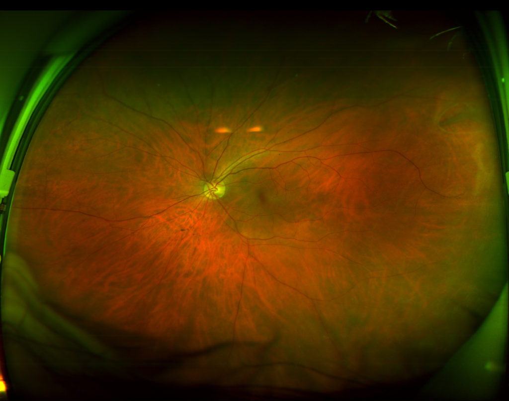
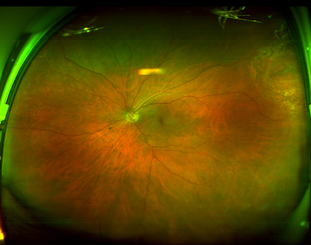
The OptoMAP retinal imaging unit has been part of our practice since 1991. This “looking in” view has become an indispensable part of a complete exam
In addition to providing extra capabilities to ascertain the “level” or “layer” of a problem, the real power in the images comes when we have annual – year after year – images to compare with each other.
The images below were from a patient with no problems in his vision. Immediately a “horseshoe tear” to the retina was observed in the right, slightly upper area (about 2:00). The patient was scheduled with a retina specialist and laser treatments were applied to prevent this from leading to a loss in vision (2nd image – after treatment).
The iWELLness retinal cross-section set of images brings detail to protection from potentially vision threatening diseases. Most appropriate for those 40 years old and above – and those younger with diabetes – this test will be added for those individuals.
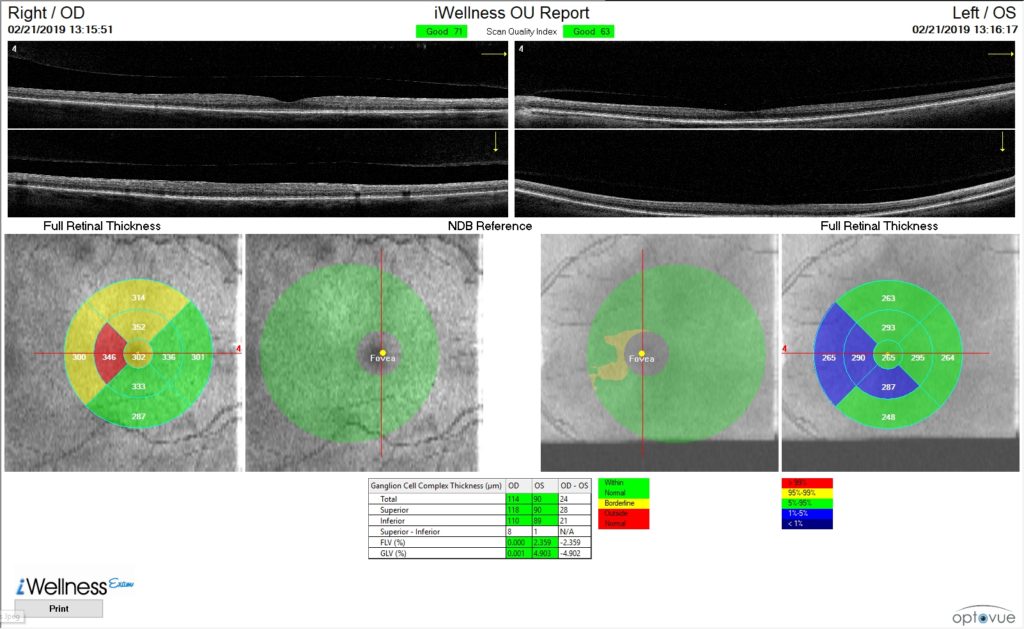 The first set of scans demonstrate a “cross-section” of the central part of our retina – the macula – your doctor will review the individual layers for thickness and regularity on direct observation.
The second set of scans demonstrate deviations from normal values. The two on the left are for the right eye of this individual, and the two on the right for the left eye. The outermost “pie charts” indicate ‘sectors’ of reduced (blue) or increased (yellow or red) thickness. The first image indicates thickening of the macula from an epiretinal membrane – not an immediate threat to vision but it will be monitored. The second and third images are comparisons to normal measurements of the “Ganglion Cell Complex” – this can begin to thin (there would be notable yellow or red areas) in glaucoma that can not be identified with traditional eye exams – this patient is normal. The fourth image shows areas of blue (thinning of the macula) which is not a specific danger to vision, but could be a potential early sign of degenerative nerve disorders (like Alzheimers Disease). ares of yellow or red indicate thickening and blue indicates a normal macula, but notable ganglion-cell-complex loss – there were NO OTHER signs of glaucoma in this patient- this screening allowed us to begin management YEARS earlier than had we waited for other glaucoma damage.
We believe that BEST CARE of your eyes includes annual retinal imaging as outlined above. The fee for these images for 2019 are:
- Optomap imaging for patients without diabetes under 40 years of age: $24
- Optomap & iWellness imaging: For those over 40 and all diabetic patients: $38